My Dog Has a Lump – Should I Panic?
My Dog Has a Lump: The Ultimate Guide to Bumps, Growths, and When to Worry
It’s a quiet evening, and you’re doing exactly what you and your dog love most: relaxing on the couch. Your hand wanders over their fur for a familiar scratch behind the ears or a belly rub, and then—your heart skips a beat. You feel it. A small, firm, or squishy protrusion that wasn’t there last week. Or was it?
The immediate internal monologue usually goes something like this: “Is it a tick? Is it a wart? Oh no, is it cancer?”
If you are currently panicking because you found a lump on your dog, take a deep breath. You are not alone, and more importantly, a lump is not an automatic death sentence. In fact, veterinary data suggests that a significant percentage of skin masses in dogs are entirely benign.
However, “wait and see” is rarely the best medical strategy. This comprehensive guide will walk you through the types of lumps dogs get, how to tell them apart, the diagnostic process, and exactly when you need to call the vet.
The “Lump Panic”: Why Our Minds Go to the Worst Case Scenario
As dog owners, our pets are family. The discovery of a growth triggers a primal fear response because we know our dogs can’t tell us if they’re in pain or if something feels “off.”
According to the American Kennel Association, roughly 20-40% of lumps found on dogs are malignant (cancerous). While those odds are high enough to warrant a vet visit, it also means there is a 60-80% chance the lump is benign.
The goal of this article is to move you from a state of panic to a state of informed action.
1. The Good News: Common Benign (Non-Cancerous) Lumps
Not every bump is a cause for alarm. Many dogs, especially as they age, develop “old dog bumps.” Here are the most common benign growths you might encounter.
Lipomas (Fatty Tumors)
The lipoma is the king of dog lumps. These are benign accumulations of fat cells.
- What they feel like: Usually soft, squishy, and movable under the skin. They are rarely painful.
- Who gets them: Middle-aged to senior dogs, and overweight dogs are more prone.
- Treatment: Usually, vets recommend leaving them alone unless they grow so large they impede the dog’s movement (like under an armpit).
Sebaceous Cysts
Think of these as the canine version of a giant pimple or an ingrown hair. They occur when a sebaceous (oil) gland becomes blocked.
- What they feel like: Firm, round, and sometimes they may have a bluish tint. If they rupture, a thick, cheesy, or oily substance might come out.
- Treatment: They may disappear on their own, or a vet may express or surgically remove them if they become chronically infected.
Histiocytomas
Often called “button tumors,” these are most common in young dogs (under 3 years old).
- What they feel like: A small, red, hairless “button” on the skin. They often appear suddenly on the head, ears, or legs.
- Treatment: These are unique because they often undergo “spontaneous regression,” meaning the dog’s immune system clears them up within 2–3 months.
Skin Tags
Just like humans, dogs get skin tags as they age.
- What they feel like: Small, fleshy stalks of skin. They are soft and usually the same color as the surrounding skin.
- Treatment: Generally ignored unless the dog keeps snagging them on things, causing bleeding.
Warts (Papillomas)
Caused by the papillomavirus, these are most common in puppies with developing immune systems or seniors with declining ones.
- What they feel like: They have a distinct “cauliflower” appearance.
- Treatment: They are contagious to other dogs but usually harmless and resolve on their own.
2. The Red Flags: Potentially Malignant (Cancerous) Lumps
While we hope for a lipoma, we must be vigilant about the “bad” lumps. Early detection is the single most important factor in a successful outcome.
Mast Cell Tumors (MCTs)
Mast cell tumors are known as “The Great Pretender” in the veterinary world. Why? Because they can look like anything. They can look like a harmless lipoma, a small wart, or a red insect bite.
- The Danger: MCTs are a form of skin cancer that can be highly aggressive or very slow-growing.
- Warning Sign: If a lump changes size rapidly (swelling and then shrinking), it could be a mast cell tumor releasing histamines.
Melanoma
While some melanomas are benign (melanocytomas), others are highly malignant.
- Where to look: Most common in the mouth, on the lips, or in the nail beds.
- Appearance: Usually dark-pigmented (black or brown) lumps.
Soft Tissue Sarcomas
These arise from connective tissues like muscle, cartilage, or nerves.
- What they feel like: Usually firm and fixed to the underlying tissue (you can’t “wiggle” them around as easily as a lipoma).
- Treatment: Often requires wide surgical excision to ensure all “tentacles” of the tumor are removed.
Mammary Gland Tumors
These occur in the mammary chain (along the belly).
- Prevention: Spaying your dog before her first heat cycle reduces the risk of these tumors to almost zero.
- The Stats: About 50% of mammary tumors in dogs are malignant.
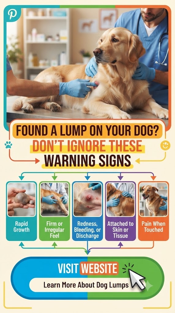
3. When is it an Emergency? (The “Red Zone” Symptoms)
While most lumps can wait for a scheduled vet appointment on Monday morning, some require immediate attention. Seek emergency care if the lump is accompanied by:
- Rapid, Explosive Growth: If a lump doubles in size in a matter of days.
- Ulceration or Bleeding: If the skin over the lump is breaking open or oozing.
- Pain or Sensitivity: If the dog yelps or snaps when you touch the area.
- Systemic Illness: If the dog has a lump AND is lethargic, losing weight, or refusing to eat.
- Interference with Function: A lump in the throat area that makes breathing/swallowing hard, or a lump on the paw that causes limping.
4. The Diagnostic Process: What to Expect at the Vet
When you take your dog to the vet for a lump, don’t expect them to just “look” at it and give you a diagnosis. Even the most experienced veterinarian cannot tell if a lump is cancerous just by feeling it.
The Fine Needle Aspirate (FNA)
This is the most common first step. The vet inserts a small needle into the lump to collect a sample of cells, which are then placed on a slide and examined under a microscope (cytology).
- Pros: Quick, usually doesn’t require sedation, and relatively inexpensive.
- Cons: Sometimes the sample is “inconclusive” because not enough cells were collected.
The Biopsy
If the FNA results are unclear, a biopsy is the next step. This involves removing a piece of the tissue (incisional biopsy) or the entire lump (excisional biopsy) while the dog is under sedation or anesthesia. The tissue is sent to a pathologist for a definitive diagnosis.
Staging and Imaging
If a lump is found to be malignant, the vet may suggest “staging.” This includes:
- Blood work: To check organ function.
- X-rays/Ultrasound: To see if the cancer has spread (metastasized) to the lungs or internal organs.
- Lymph Node Aspirates: To check if the cancer has moved into the lymphatic system.
5. Factors That Influence Risk
Breed Predisposition
Genetics play a massive role in whether a dog develops lumps.
- Boxers, Beagles, and Boston Terriers: Highly prone to Mast Cell Tumors.
- Golden Retrievers: Unfortunately, they have high rates of various cancers, including Hemangiosarcoma and Lymphoma.
- Dachshunds: Prone to fatty lipomas.
- Schnauzers: Often develop “Schnauzer bumps” (comedone syndrome).
Age
As dogs age, their cell replication processes become less “perfect,” leading to more errors and more growths. It is common for a 10-year-old dog to have half a dozen various bumps.
6. How to Perform a “Monthly Lump Check” (The Lump Map)
Prevention is difficult, but early detection is easy if you are proactive. Once a month, perform a “nose-to-tail” exam on your dog.
- Start at the Nose: Check the muzzle and gums.
- Feel the Ears: Run your fingers along the flaps and the base.
- The Neck and Shoulders: Feel deep into the fur.
- The Legs and Paws: Check between the toes and around the nail beds.
- The Belly: Feel along the mammary chain.
- The Tail and Perianal Area: Don’t skip the “underneath” parts.
Creating a “Lump Map”
If you find a lump, start a “Lump Map.” Draw a basic outline of a dog on a piece of paper and mark the location of the lump. Write down:
- Date discovered.
- Size (Use a ruler or compare it to a coin/pea).
- Texture (Hard, soft, movable).
- Appearance (Red, skin-colored, hairless).
This map is incredibly helpful for your vet to track changes over time.
7. Treatment Options: Moving Forward
If the diagnosis comes back and it’s something that needs to be addressed, you have several paths.
Surgical Removal
For many skin tumors, surgery is curative. If the vet can get “clean margins” (meaning they remove the tumor and a healthy ring of tissue around it), the problem may be solved forever.
Chemotherapy and Radiation
If the tumor is aggressive or has spread, veterinary oncologists can use chemotherapy or radiation. Unlike human chemo, dog chemo is designed for quality of life first. They usually don’t lose their hair or get as sick as humans do; the goal is to manage the disease, not necessarily “cure” it at the cost of the dog’s happiness.
The “Watch and Wait” Approach
If the lump is a confirmed benign lipoma and it’s not bothering the dog, the best treatment is often no treatment at all. Surgery carries risks (anesthesia, infection), so if a lump is harmless, we leave it be.
Holistic and Supportive Care
While not a replacement for medical treatment, many owners use anti-inflammatory diets, supplements like Turmeric/Curcumin (with vet approval), and high-quality Omega-3 fatty acids to support the immune system of a dog dealing with growths.
8. Managing the Emotional Toll
Finding a lump on your dog is stressful. It’s okay to feel upset. However, remember that veterinary medicine has advanced leaps and bounds in the last decade. Many conditions that were once terminal are now manageable or curable.
The best thing you can do for your dog is to remain calm. Dogs are experts at reading our energy. If you are frantic, they will be stressed. Treat the vet visit as just another “adventure” or errand, and provide lots of treats and praise.
9. Common Myths About Dog Lumps
Myth #1: “If it doesn’t hurt the dog, it’s not cancer.” Reality: Many malignant tumors are completely painless in the early stages. Pain is not a reliable indicator of danger.
Myth #2: “If it’s soft, it’s just fat.” Reality: While lipomas are usually soft, some Mast Cell Tumors can also feel soft and squishy. Never assume based on texture alone.
Myth #3: “He’s too young for cancer.” Reality: While rare, young dogs can develop aggressive tumors. Age makes lumps more likely, but it doesn’t make a young dog immune.
Myth #4: “If I poke it (FNA), it will cause the cancer to spread.” Reality: This is an old myth. The risk of “seeding” cancer cells via a fine needle is extremely low and far outweighed by the benefit of knowing what the lump is.
10. Summary Checklist for Owners
If you just found a lump, follow these steps:
- Don’t Panic: Remember the 60-80% rule for benign growths.
- Measure it: Get a baseline size.
- Check for “Red Flags”: Is it bleeding? Is the dog acting sick?
- Schedule a Vet Visit: Even if it feels like a “fatty tumor,” get a professional opinion for peace of mind.
- Ask for an FNA: Ensure the vet takes a cell sample rather than just “eyeballing” it.
- Update your Lump Map: Keep track of it for the rest of the dog’s life.
Conclusion: Knowledge is the Best Medicine
Your dog relies on you to be their advocate. By noticing that lump, you’ve already done the hardest part of being a vigilant pet parent. Whether it turns out to be a harmless “old dog wart” or something that requires surgery, catching it early gives your dog the best possible chance at a long, happy, and waggy-tailed life.
So, put down the search engine, pick up the phone, and call your vet. Once you have a plan in place, the panic will fade, and you can get back to what really matters: those belly rubs and couch snuggles.
Disclaimer: This article is for informational purposes only and does not constitute medical advice. Always consult with a qualified veterinarian regarding your pet’s health.